Lower Extremity Vein Mapping Ultrasound Protocol – This systematic review aims to assess relationships between lower limb peripheral venous diseases (lower limb varicose veins (LLVV), venous thromboembolism (VTE) comprising deep vein thrombosis and . Characterization and follow-up of deep vein stents can be achieved by ultrasound and histology. This viable animal model can identify human pathological stent processes as early thrombosis, resolution .
Lower Extremity Vein Mapping Ultrasound Protocol
Source : www.researchgate.net
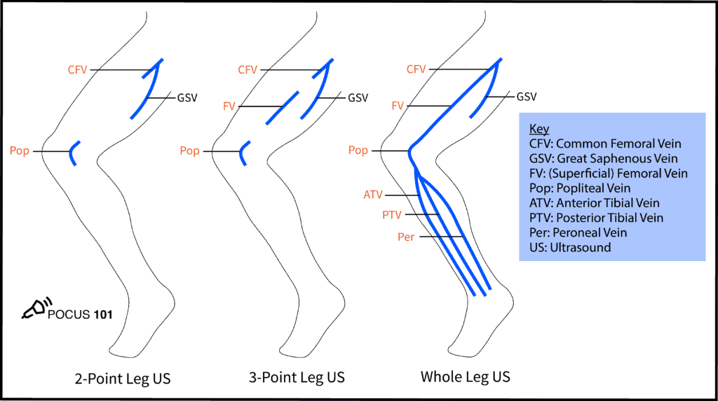
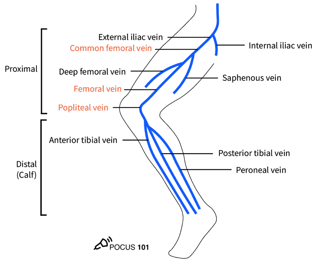
DVT Ultrasound Made Easy: Step By Step Guide POCUS 101
Source : www.pocus101.com
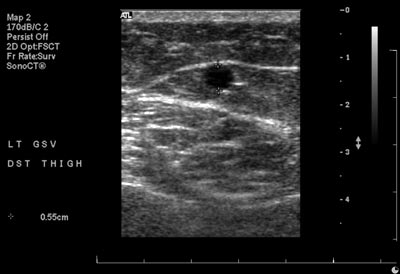
Case study 2: duplex ultrasound mapping scan. Shaded veins
Source : www.researchgate.net
Lower Extremity Venous Protocols and Interpretation YouTube
Source : www.youtube.com
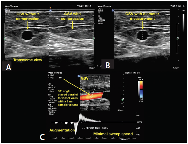
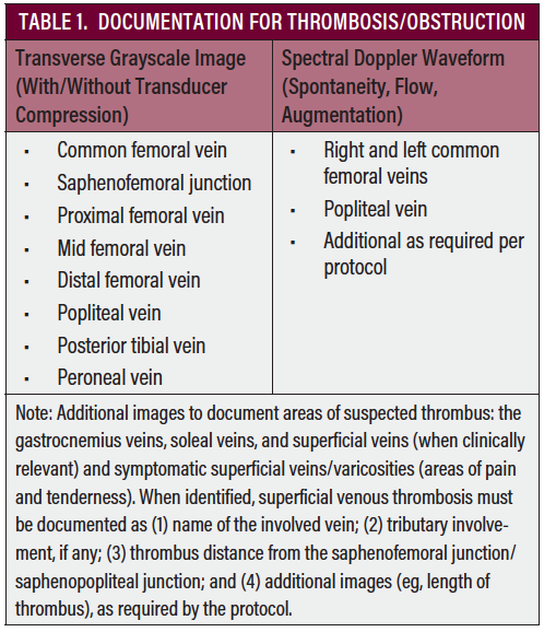
Duplex Ultrasound Technical Considerations for Lower Extremity
Source : evtoday.com
DVT Ultrasound Made Easy: Step By Step Guide POCUS 101
Source : www.pocus101.com
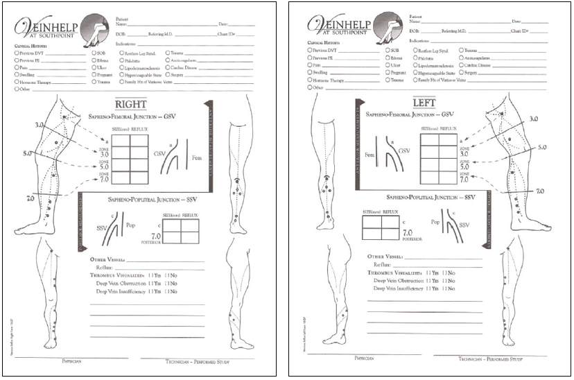
Vein Mapping | Vascular Center | UC Davis Health
Source : health.ucdavis.edu
Duplex Ultrasound Technical Considerations for Lower Extremity
Source : evtoday.com
Example of the schematics of a lower extremity vein mapping for
Source : www.researchgate.net
Duplex Ultrasound Technical Considerations for Lower Extremity
Source : evtoday.com
Lower Extremity Vein Mapping Ultrasound Protocol Example of the schematics of a lower extremity vein mapping for : Objective: To design, implement and assess a clinical pathway for lower-extremity deep venous thrombosis, and to compare the length of hospital stay in two different periods. Design: Development of . Ultrasound is not necessary when computed or D-dimer measurement plus bilateral lower extremity venous compression US (if D-dimer level is >500 ng/mL), followed by chest CT if US results .

